Abstract
Age-related macular degeneration is a degenerative disorder of the macula which is characterised, in its early stages, by a partial sensitivity loss to contrast particularly at mesopic and scotopic light levels and a slower dark adaptation mechanism. Metropsis offers an easy and quick contrast sensitivity test that can be run at these crucial light levels.
Keywords: AMD, age-related macular degeneration, dry-AMD, wet-AMD, contrast sensitivity, CSF, Metropsis, photoreceptor, scotopic, mesopic, visual function, cones, rods, children, paediatric, visual function, aging, adaptive procedure, psychophysics, spatial frequency.
What is AMD?
Age-related macular degeneration (AMD) is a degenerative disorder of the central retina, the macula. Early signs of AMD consist of morphological changes in the macula which tend to appear in people over the age of 50 years. A proportion of these patients develops a severe and irreversible loss of central vision in one or both eyes. The macular dysfunction is often due to either an extensive atrophy of the tissue – also known as geographic atrophy (GA) or dry-AMD – or because of scarring due to a neovascular lesion – also known as choroidal neovascularization (CNV) or wet-AMD.
Why is it so difficult to detect the onset of AMD?
It is not easy to measure early functional changes that occur in AMD. This is because early changes are typically of a morphological nature and occur with unnoticeable vision loss or measurable visual dysfunction. These changes need to become much more pronounced for the patient to be aware of correlated functional vision changes, such as difficulty in reading under dim light and problems with dark adaptation.
These changes can also be missed by conventional vision tests used in the clinic, such as the distance visual acuity tests1. This is probably because the visual acuity changes are too small to be significantly different from the test-retest reliability of the acuity charts2,3,4. Also, these eye charts are limited (i) to one level of contrast, (ii) to a high spatial frequency content of the letters presented, and (iii) to the assessment of the central visual field.
What is the best way to measure early functional vision changes?
Tests that measure the sensitivity to contrast at different spatial frequencies provide more information about the functionality of the photoreceptors and their postreceptoral pathways. These tests are being used more and more often to quantify functional vision loss in AMD.
Tests that measure the sensitivity to contrast at different spatial and temporal frequencies provide more information about the functionality of the photoreceptors and their postreceptoral pathways. These tests are being used more and more often to quantify functional vision loss in AMD.
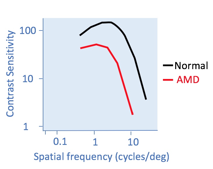
It has been demonstrated that the contrast sensitivity function of AMD patients is similar in shape to the normal CSF, but it is shifted along the spatial frequency and the contrast sensitivity axes5. The figure below (redrawn from Chung and Legge, 2016) illustrates this shift.
Go to more information on the spatial contrast sensitivity tests available with Metropsis.
Learn how to test spatial contrast sensitivity at different light levels.
Go to more information on the temporal contrast sensitivity tests available with Metropsis.
References
2Rosser, D.A., Cousens, S.N., Murdoch, I.E., Fitzke, F.W., Laidlaw, D.A., 2003. How sensitive to clinical change are ETDRS logMAR visual acuity measurements? Invest. Ophthalmol. Vis. Sci. 44, 3278–3281.
3Arditi, A., Cagenello, R., 1993. On the statistical reliability of letter-chart visual acuity measurements. Invest. Ophthalmol. Vis. Sci. 34, 120–129.
4Lovie-Kitchin, J.E., Brown, B., 2000. Repeatability and intercorrelations of standard vision tests as a function of age. Optom. Vis. Sci. 77, 412–420.
5Chung, S.T., Legge, G.E., 2016. Comparing the Shape of Contrast Sensitivity Functions for Normal and Low Vision. Invest. Ophthalmol. Vis. Sci. 57, 198-207.