Abstract
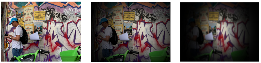
Peripheral vision is crucial for navigating through the environment and planning where to look when performing a visual search. This ability appears to be severely affected in retinitis pigmentosa and glaucoma patients, for whom the loss of peripheral vision often precedes the loss of foveal vision (Figure 1). Patients who have suffered from stroke can also show intact foveal vision, while demonstrating hemianopia or cortically-induced blindness in the visual field contralateral to the lesion.
A way to assess vision in the periphery is by testing peripheral contrast sensitivity. This test measures the ability to discriminate a pattern that appears at different locations across the visual field. Areas of low sensitivity will be associated with areas of vision loss.
We have developed a Peripheral Contrast Sensitivity Function (CSF) Test that allows investigators to measure contrast sensitivity only at selected locations in the visual field, thus saving testing time.
Keywords: contrast sensitivity function, CSF, peripheral vision, glaucoma, retinitis pigmentosa, hemianopia, perimetry, eye tracker.
Why measure peripheral contrast sensitivity?
Peripheral vision is typically intended as the area of the visual field outside the parafovea or the central 4°-5° around the fovea1.
Some visual deficits, like glaucoma and retinitis pigmentosa, may affect peripheral vision before they impact foveal vision, others show a rate of progression which is faster in the periphery than in the fovea. In these cases, measurements of foveal contrast sensitivity cannot be extrapolated, thus peripheral vision needs to be measured directly.
How can I measure peripheral contrast sensitivity?
To measure contrast sensitivity in the periphery, observers are asked to fixate a cross in the centre of the screen while a test pattern (grating) appears at eccentric locations. The spread and number of tested locations varies from test to test which directly affect the test duration.
How can I measure peripheral contrast sensitivity with Metropsis?
The Peripheral Contrast Sensitivity Function Test implemented in Metropsis presents gratings only at targeted locations across the visual field which are preselected by the examiner. In this way, investigators can concentrate on monitoring changes in vision only at locations that have been deemed to be of interest. This option dramatically reduces the testing time and thus the quality of the data collected.
During the Peripheral Contrast Sensitivity Function Test, the observer is instructed to fixate a cross displayed in the centre of the screen and respond to the orientation of the grating that appears in the periphery (Figure 2).
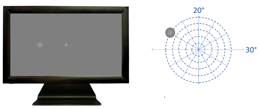
Right: Schematic representation of possible stimulus locations (dotted lines).
To make sure that we are measuring contrast sensitivity at the intended location, it is crucial to monitor fixation. Metropsis uses an eye tracker (Figure 3) to check that the observer is fixating; if the observer is not fixating, the trial is discarded.
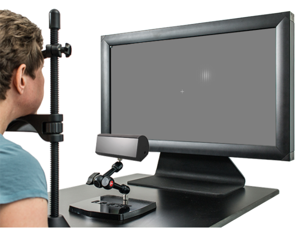
Figure 4. Presentation of a stimulus (top) and output from LiveTrack FM eye tracker (bottom).
Why is the Metropsis Peripheral Contrast Sensitivity Test better than other commercially available tests?
- Peripheral vision is typically assessed during a perimetry examination. However, perimeters often are not sensitive enough to detect small spontaneous visual deteriorations or artificially induced visual improvements. The Metropsis Peripheral CSF Test measures the minimum amount of contrast required to discriminate the orientation of a grating. This task is more challenging than a detection task used by perimeters and thus more sensitive to reveal small changes in visual thresholds.
- Metropsis uses custom-designed display technology with integrated calibration to deliver accurate visual stimuli. It provides very fine control of colour and contrast, which allows the examiner to detect subtle changes in visual functions.
- The Metropsis Peripheral CSF Test allows to test sensitivity of only targeted locations of the visual fields that are specified by the investigator, saving time from testing uninformative locations.
- The Metropsis Peripheral CSF Test uses an eye tracker to monitor fixation, which is more reliable than using negative responses of the blind spot to assume fixation.
- The Metropsis Peripheral CSF Test can be used for visual training of targeted areas of interest.
- The Metropsis Peripheral CSF Test can also be run at different light levels, such as mesopic or scotopic.